VO-CRO In Vitro Cell Types
VO-CRO scientists have a collective 80 years of experience isolating and culturing multiple ocular cell types from a variety of species including mouse, rat, and human. Because our focus is on providing the highest quality and most clinically relevant and translatable research possible, the use of primary human cells is very important to us. We use a wide variety of ocular cells in order to determine the relative contribution of each cell type in eye disease onset, progression, and regression.
CELL TYPES

HUMAN

RAT
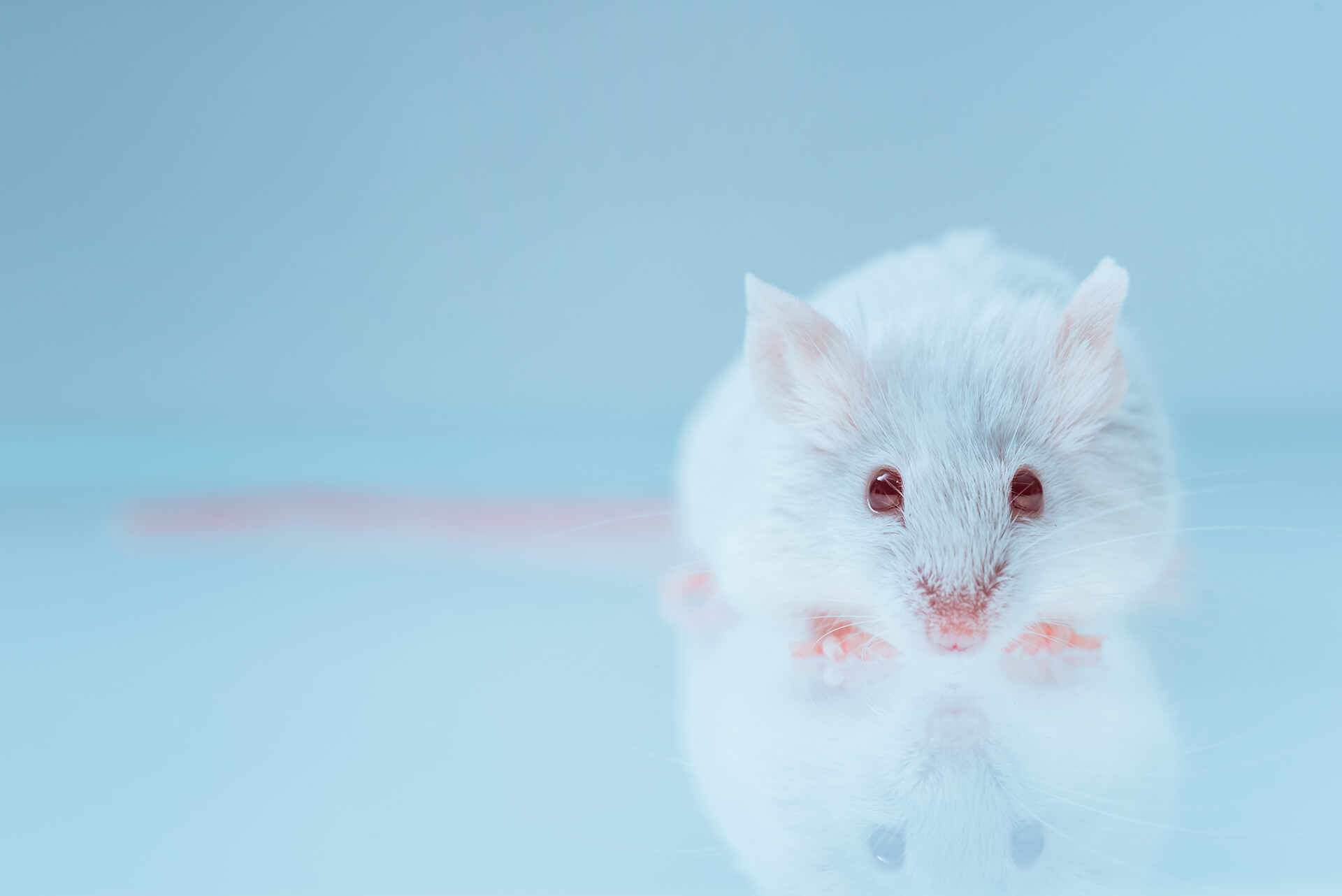
MOUSE
Choroidal Endothelial Cells
The choroid is a fenestrated vascular bed that provides nutrients to the photoreceptors. These cells are particularly important in the pathogenesis of age-related macular degeneration. These cells can be treated with disease-relevant stimuli to induce expression of cytokines and growth factors allowing for the careful examination of pro-inflammatory and pro-angiogenic signaling pathways.
Retinal Microvascular Endothelial Cells
Retinal Microvascular Endothelial Cells (RMEC) comprise the microvascular network of the inner retina. Their primary role is to provide nutrients to retinal cells while maintaining the blood-retina barrier. Like choroidal endothelial cells, retinal microvascular endothelial cells can be stimulated to express gene products that are important in disease pathogenesis. Using in vitro methods, the molecular pathways involved in disease pathogenesis can be examined under tightly controlled experimental conditions.
Retinal Pericytes
Pericytes are the support cells of the vasculature. Normal pericyte function is critical to the survival of microvascular endothelial cells under pathologic conditions and appropriate regulation of vascular permeability. Human pericytes can be cultured to examine the regulation of apoptotic pathways, cytokine production and other cell behaviors leading to disease onset and progression
Müller Glia
Müller cells are specialized glial cells that nearly span the thickness of the retina. They are the retina’s primary producers of cytokines and growth factors. For example, Müller cells produce many-fold more VEGF, TNF-alpha, interleukins and other growth and inflammatory factors than all other retinal cell types combined. We culture these cells to explore the signal pathways and intermediates that regulate the production of these and other factors under disease-relevant conditions.
At VO-CRO, our focus is on providing the best and most translatable research possible.
Contact Us To Learn MoreRetinal Pigmented Epithelial Cells
Retinal Pigmented Epithelial Cells (RPE) is a layer of pigmented epithelial cells responsible for the permeability barrier between the retina and the choroid. RPE are also important sources of growth factors and cytokines that promote disease progression. We culture and study RPE in these two contexts, using TEER, ELISA, qRT-PCR and other methods.
Astrocytes
Retinal astrocytes help to maintain retinal homeostasis. Because astrocytes can express and secrete growth factors and cytokines, they are among the battery of cells with which we examine these functions under disease-relevant conditions. Our overall purpose is to determine the relative contributions of the various retinal cell types in disease onset, progression, and regression.
Microglia
Microglia are a type of glial cell found in the inner layers of the retina. As the resident immune cells of the retina, microglia contribute to the inflammatory response by secreting tumor necrosis factor-α (TNF-α) and interleukin-1β (IL-1β), two cytokines implicated in several retinal diseases. Additionally, retinal microglia may influence the branching and expansion of the retinal vasculature, so they are studied closely in relation to vasculogenesis and angiogenesis.
Neuro
Retinal neurons include rod and cone photoreceptors, bipolar cells, horizontal cells, amacrine cells, and ganglion cells. Together these form the retina’s neuronal circuits, which produce signals that are sent to the brain for visual processing. As terminally differentiated cells, retinal neurons can only be maintained in culture for a limited time. VO-CRO has optimized our methods to allow for investigations of these cells’ behaviors within the period of cell viability in culture.
Want to know more? Visit our Readouts page or Contact our team.